




















Laboratory diagnostics: <em>E. coli</em> (diarrhea)
What laboratory diagnostic methods can I use to diagnose E. coli? Which one should I choose according to the situation? How do I interpret the results?

Assays available
Bacterial culture
- Isolation of live organism.
- Sample types: feces, intestinal content.
- Pros:
- Bacteria is easy to grow in 1 day.
- Easy to do in any lab (including in-house).
- Relative low cost.
- Cons:
- Some E. coli are part of normal intestinal flora.
- Pigs have multiple E. coli at same time.
- Pigs previously treated with antibiotics can prevent bacterial growth.
Antimicrobial susceptibility
- Tests in vitro ability of live organism to grow under specific concentrations of different antimicrobials.
- Sample types: feces, intestinal content.
- Pros:
- Identification of susceptibility or resistance of specific isolate to common antimicrobials.
- Identification of antimicrobial resistance trends.
- Cons:
- Requires a bacterial isolate.
- In vitro testing may be slightly different than in vivo results.
- Some specific antimicrobials may not be tested or require separate, special testing.
- Moderate cost.
Histopathology
- Evaluates the presence of tissue lesions (damage) which can confirm the presence of disease. Sometimes can also detect the presence of organisms directly (bacteria and parasites) or indirectly (virus) through additional special staining.
- Sample types: tissue.
- Pros:
- Associate bacterial adhesion with intestinal damage.
- No special staining needed.
- Cons:
- Need to formalin-fix intestinal tissues within <15 min of pig death -Intestinal tissue decomposes quickly.
- Disease can be segmental so multiple intestinal samples should be submitted.
Genotyping
- A polymerase chain reaction (PCR) technique that detects presence of specific sequence of nucleic acids (DNA) associated with known virulence genes.
- Pros:
- Done via PCR; easy to do.
- Helps identify pathogenicity by detecting presence of genes (not necessarily expression of genes) associated with virulence factors.
- Toxin Genes: EAST1, LT, STa, STb, Stx1, Stx2, Stx2e.
- Pilus Genes: F18, F41, K88 (F4), K99 (F5), 987P (F6).
- Adhesin Genes: AIDA, EAEA, PAA.
- Helps with proper vaccine selection (matching correct pili).
E. coli genes detected though PCR genotyping and their function.
| Gene | Type | Description |
|---|---|---|
| EAST1 | Toxin | Intestinal toxin binds to the same receptor and stimulates the same secretory pathway as Sta (causation of diarrhea is not well established) |
| LT | Toxin | Intestinal toxin that stimulates intestinal secretion |
| STa | Toxin | Intestinal toxin that stimulates intestinal secretion mostly in young pigs (decreases absorption of water an electrolytes) |
| STb | Toxin | Intestinal toxin stimulating secretory diarrhea mostly in older pigs (increases secretion from enterocytes) |
| Stx1 | Toxin | Intestinal toxin can be found in pathogenic and non-pathogenic strains |
| Stx2 | Toxin | Intestinal toxin can be found in pathogenic and non-pathogenic strains |
| Stx2e | Toxin | Systemic toxin which induces vascular damage and edema disease |
| F18 (F107) | Adhesin | Pili (fimbria) used for attachment which is present after 20 days of age and commonly associated with post weaning scours and edema disease |
| F41 | Adhesin | Pili (fimbria) used for attachment mostly neonates |
| K88 (F4) | Adhesin | Pili (fimbria) used for attachment birth through all ages |
| K99 (F5) | Adhesin | Pili (fimbria) used for attachment mostly neonates |
| 987P (F6) | Adhesin | Pili (fimbria) used for attachment mostly neonates |
| AIDA | Adhesin | Nonfimbrial “adhesin-involved-in-diffuse-adherence” more commonly found in conjunction with F18 |
| EAE | Adhesin | Nonfimbrial adhesin that contributes to "attaching-and-effacing" lesions |
| PAA | Adhesin | Nonfimbrial adhesin essential for producing "attaching-and-effacing" lesions |
- Cons:
- Requires a bacterial isolate.
- Hope the correct isolate is genotyped (pigs can have several isolates at the same time).
- Moderate price, but usually only test 1 isolate.
Result interpretation
Bacterial culture
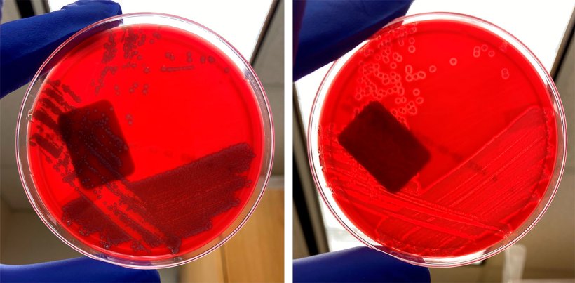
- Interpretation of hemolysis depends on pigs age:
- Pre-wean: non-haemolytic E. coli.
- Post-weaning: haemolytic E. coli.
- Purity:
- Pure growth: highly suggestive of disease contributor.
- Mixed growth: questionable value.
- Amount:
- High: highly suggestive of disease contributor.
- Moderate: variable interpretation.
- Low: questionable value (could be contaminant).
- No growth: Animal possibly previously treated with antibiotics or not significant contributor.
Antimicrobial susceptibility
- Susceptible: possible good choice for treatment if antimicrobial can reach target tissue.
- Resistant: select different antimicrobial
- MIC: if MICs are done ensure antimicrobial selected achieves the listed MIC value in the target organ.
Histopathology
- Positive: Strong association with accusation if able to demonstrate intestinal lesions with E. coli adhered to epithelial cells.
- Negative: No intestinal lesions.
Genotyping
- Genotyping is very valuable in identifying if isolated E. coli contains any of the virulence factors that would support the isolate as pathogenic.
Scenario
Pigs with scours (any age):
- Collect rectal swabs from 2 or more untreated scouring pigs and submit for bacterial culture, antimicrobial susceptibility, and genotyping.
- Euthanize 1-3 untreated scouring piglets. Immediately collect and place in formalin solution 3 intestinal samples from each piglet. Collect fresh intestinal samples to submit chilled for bacterial culture, antimicrobial susceptibility, and genotyping.